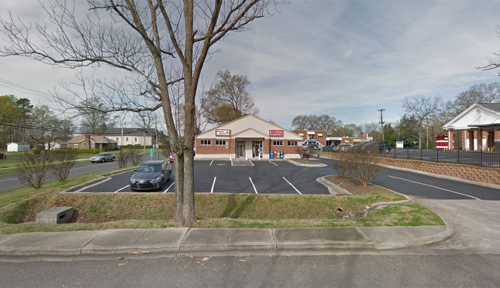
Dallas Express Pharmacy
111 A North Hoffman Street
Dallas, NC 28034
(704) 922-3001
Mon-Fri 8:30am - 6:00pm
Sat Closed
Sun Closed
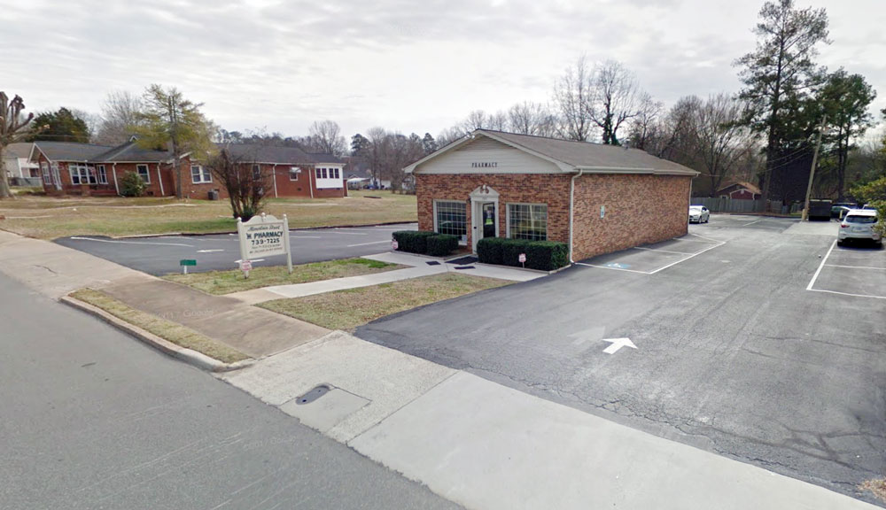
Mountain Street Pharmacy
709 West Mountain Street
Kings Mountain, NC 28086
(704) 739-7225
Mon-Fri 8:30am - 6:30pm
Sat Closed
Sun Closed
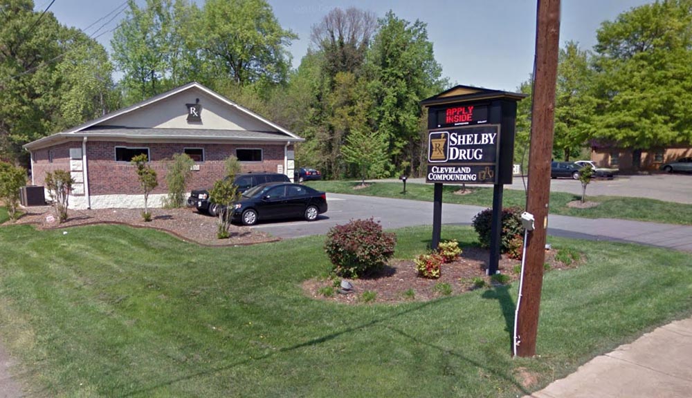
Shelby Drugstore
701-1 E. Grover St.
Shelby, NC 28150
(704) 487-2939
Mon-Fri 8:30am - 6:30pm
Sat 8:30am - 12:30pm
Sun Closed